
Veterinary medicine is not just black and white; it’s all shades of grey!
As I sit here writing this blog post and thinking back on how amazing my first week at my externship has been, I just can’t believe how much I have experienced already! I am very fortunate to be spending the months of May and June with New Hamburg Veterinary Clinic because of the amazing mentorship they provide and the vast amount of clientele they serve. Each day has been a new adventure and every morning I wake up with more energy than I ever have had in anticipation of the day that is to come. To give you a quick overview of just how busy a day can be at the clinic, my Friday consisted of pregnancy checking Standardbred mares via ultrasound, routine inspection of a guinea pig farm that supplies major pet stores globally, caring for an in-hospital canine patient with MODs (multi-organ dysfunction), and dehorning a group calves using techniques such as thermal cauterization. In just one week it seems like I have learned and practiced more clinical skills than I have in the past three years!
One of the greatest lessons I have learned this week is how valuable utilizing diagnostic imaging can be as a tool in working up a case. It is a great preliminary diagnostic test to gain a general overview of how the organs are looking, and thus functioning, in the body. The clinic is very fortunate to offer in-house digital radiography, dental radiography, and ultrasound and this equipment is used every day by the veterinarians to provide the greatest possible care for their patients. Diagnostic imaging has historically been very intimidating to me. We have studied diagnostic imaging in school for the past three years, but as a student I have a hard time interpreting radiographs in a classroom setting without the background knowledge of a clinical case. Radiology is a skill set that needs to be learned through repetition and pattern recognition, and with little experience interpreting radiographs “in the real world” I was nervous for when the time came to do so during my externship. Luckily for me, the veterinarians are all very willing to take the time to describe what they are seeing and have helped me better understand the 50 shades of grey that I see on the screen.
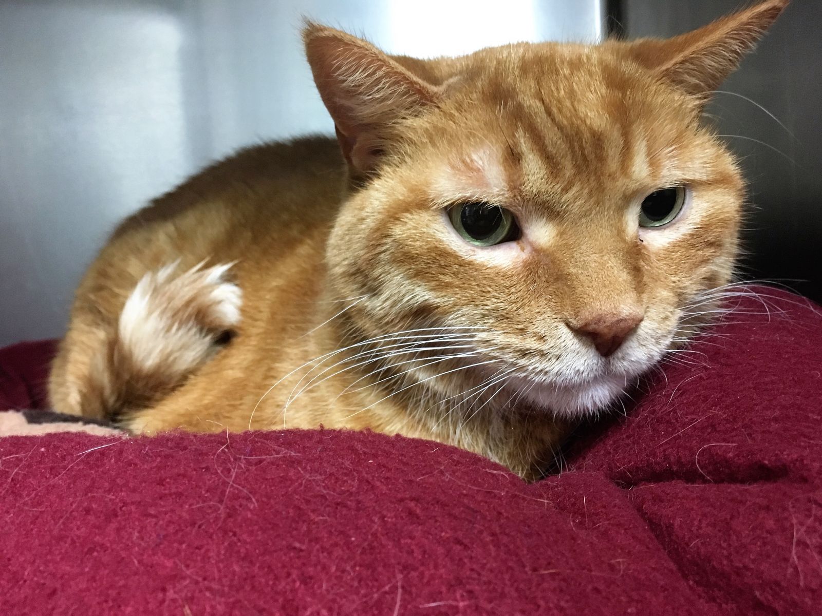 Lester B. Pearson, or Lester for short, is one such patient who came into the clinic this week that had diagnostic imaging as one of the diagnostic tests performed for his case. Lester is a very handsome mature, neutered male, orange tabby cat who belongs to one of the vets. He was brought into the clinic for lethargy, vomiting, sneezing, and chronic weight loss. His mum was worried that there could be an underlying problem to explain all of these symptoms and brought him in to have a panel of tests performed. His SDMA blood levels were elevated, which is indicative of Stage 1 kidney disease. Kidney disease is not uncommon in older feline patients, so the next step was to take a look at Lester’s thorax and abdomen to see if there was anything else going on that could also explain his symptoms.
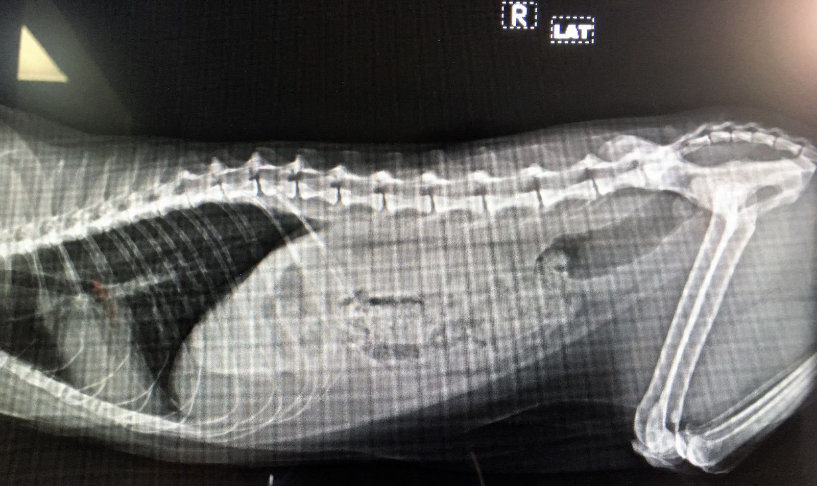
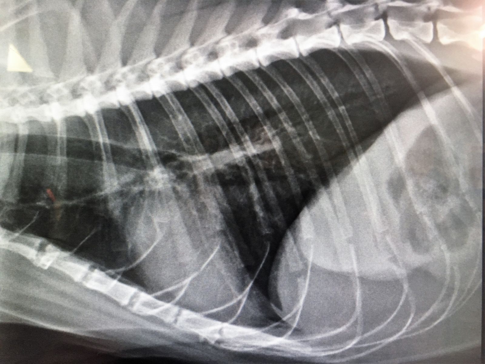
A full body radiograph was performed to get a generalized picture of how his internal organs appear. In the first radiograph above we can see that Lester’s kidneys are a normal size which supports the diagnosis of only Stage 1 kidney disease. An indication of more advanced chronic renal disease would be kidneys that are small in size and/or irregularly shaped. The rest of his abdominal organs look normal and nothing seemed to jump out at us that would indicate a cause for his symptoms. Having an unremarkable radiograph is not a bad thing; it’s what we hope for! This means that Lester does not have significant organ dysfunction that would show up with radiographic imaging. Despite a normal abdominal radiograph, a small pulmonary nodule was noted on the thoracic half of the radiograph (second radiograph above). The nodule was only seen on the right lateral view and could just be due to his age. Can you spot the nodule?
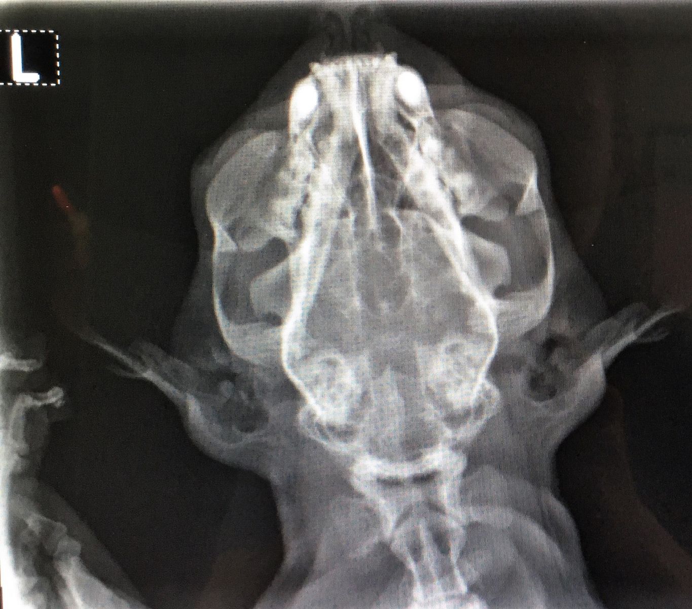 The pulmonary nodule in Lester’s lungs does not quite explain the sneezing fits he has once a day. A head radiograph was also performed to see if there could be anything blocking and causing irritation to his nasal cavity and sinuses. Based on the radiograph there does not appear to be anything physical, such as a tumour or foreign body, causing the sneezing. His mum’s next step in terms of diagnosing a cause of the sneezing is to perform a nasal flush for cytology under general anesthesia when he gets a dental cleaning done in a couple of months.
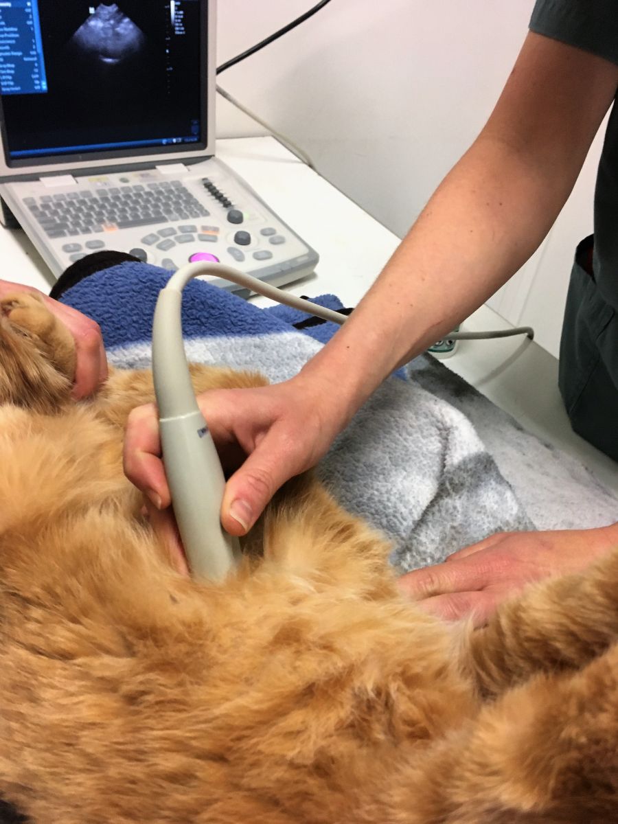
As mentioned above, the clinic is very fortunate to have an in-house ultrasound that can be used for additional diagnostics if the radiographs come back inconclusive. Ultrasound is a great diagnostic tool that uses soundwaves to produce a real-time image of the internal organs, which is both a safer and more sensitive test than radiography. In the photo below you can see Lester’s mom performing a general scan of his abdomen to see if there is anything blaringly apparent that was missed by radiographs. Again, his kidneys appeared to be normal in size and shape and no abnormalities were noted. Based on both the unremarkable radiographs and ultrasound it is safe to say that Lester has no major concerns going on right now and his vomiting can be attributed to some inflammation in his bowels. His mum has switched him to a GI diet and I am happy to report that Lester has not vomited since!
 I cannot wait to continue practicing my diagnostic imaging skills and having the confidence to use this skill-set while working up a case in the clinic. Although some patients may have easily diagnosable conditions via imaging, others may just need imaging to rule out disease; this is why veterinary medicine is not always a ‘black and white’ science!