This week, I spent time reflecting on some of the amazing things I have learned during my externship here on Vancouver Island. It brought me to thinking about just how lucky we are to be in a profession where we are constantly challenged; constantly faced with changing science and a new standard of doing things. And, with this reflection, I began to see that those who accept the ever-progressing field of medicine and work towards always improving their skills, learning from challenging cases, or recognizing where they have made mistakes to do better in the future, are the practitioners who have the most satisfying careers. It is these types of everyday challenges in a career of veterinary medicine that makes it so exciting! What an incredibly mentally, emotionally, and often physically stimulating career.
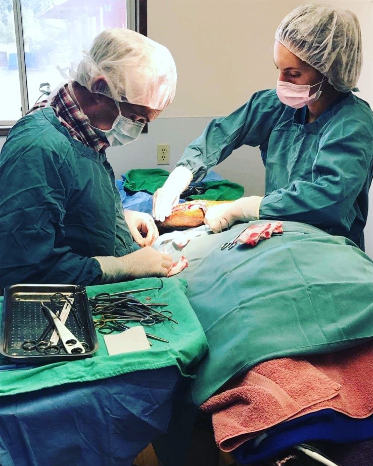
 I was lucky enough this week to scrub in on a tightrope surgery at Comox Valley Animal Hospital with Dr. Dave MacDonald. This is one of several times I have been able to participate in this procedure here at the clinic. However, prior to coming out on the island, I had never even seen this procedure performed. So naturally, I was excited to expand my knowledge and skill set!
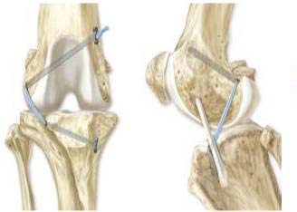
At referral centres, like the OVC, board certified specialists perform Tibial Plateau Leveling Osteotomy (TPLO) or the Tibial Tuberosity Advancement surgery (TTA) (based on surgeon’s preference and expertise) instead of the tightrope procedure because of a higher success  rate post-operation. Both the TPLO and TTA surgeries involve changing the anatomy and thus mechanics of the bones involved in the stifle (knee) joint, and are performed to stabilize the joint after ruptures of the cranial cruciate ligament (analogous to the anterior cruciate ligament in humans). In contrast, the tightrope surgery (shown on right) is an extracapsular technique where heavy polyester sutures are placed through holes drilled in the tibia and femur to physically try and mimic the ruptured cranial cruciate ligament.
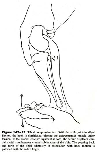
The cranial cruciate ligament is responsible for stifle stabilization, and attaches the caudal aspect of the femur (back portion) to the cranial aspect of the tibia (front portion), preventing the tibia from sliding forward. It is unknown exactly why the cranial cruciate ligament ruptures in dogs, but the reasoning is perceived to be multifactorial in nature. Genetic predisposition, repetitive stress on the stifle joint, excess weight, and trauma are all thought to play a role. Often, there is a chronic, progressive weight bearing lameness on the affected leg, and rupture occurs from an acute minor trauma (but the disease process has been there a long time). Unfortunately, when one cruciate ruptures, it is much more likely that the second leg will also rupture at some point in time. Large, middle aged dogs (~4 years old) and older smaller breed dogs (>7years old) tend to be the most commonly affected for this type of tear. The injury can occur in cats, but it is uncommon. Diagnosis is made by the veterinarian using a combination of clinical signs, history from the owner, and the cranial drawer/tibial compression tests (shown below).
.png)

Although the treatment of choice for dogs over 10 kg is the TPLO or TTA procedure, based on success rate post-operatively, this is often not a financially viable option for many dog owners here and includes a long trip to a referral centre hours away. In comparison, the tight rope procedure offered at this clinic is relatively less expensive, and thus the only viable surgical option for affected dog owners. For this reason, it was very interesting to learn about the tightrope surgery, and watch it being performed in a general practice setting. One more new and exciting thing I have learned during my externship here!
Illustrations from: “Cranial Cruciate Ligament Rupture” Dr. Noël Moens, VETM 4470, University of Guelph, Ontario, Canada, Presented February 6, 2017.
