Hi everyone! In week five, I learned how to perform a new surgery, got to practice my orthopedic and neurologic exams, and got to see a pretty dramatic case (I have pictures!).
Let’s start with the new surgery. In school, one of our lectures included a condition called an “aural hematoma”. It’s a relatively simple condition, but one I figured I wouldn’t see often in practice. Was I ever wrong! At this point in the summer, I’ve already seen about five or six dogs come in with aural hematomas.
An aural hematoma results when the cartilage and blood vessels of the ear become damaged, leading to a painful accumulation of blood between the two layers of cartilage that form the ear flap (pinna). This typically occurs when a dog has been excessively shaking its head or scratching its ears, due to an underlying cause such as an ear infection, allergies, or ear mites. If left untreated, the blood slowly drains out, but leaves lots of scar tissue, and becomes what is commonly known as “cauliflower ear” – a slightly shrunken, crinkly ear.
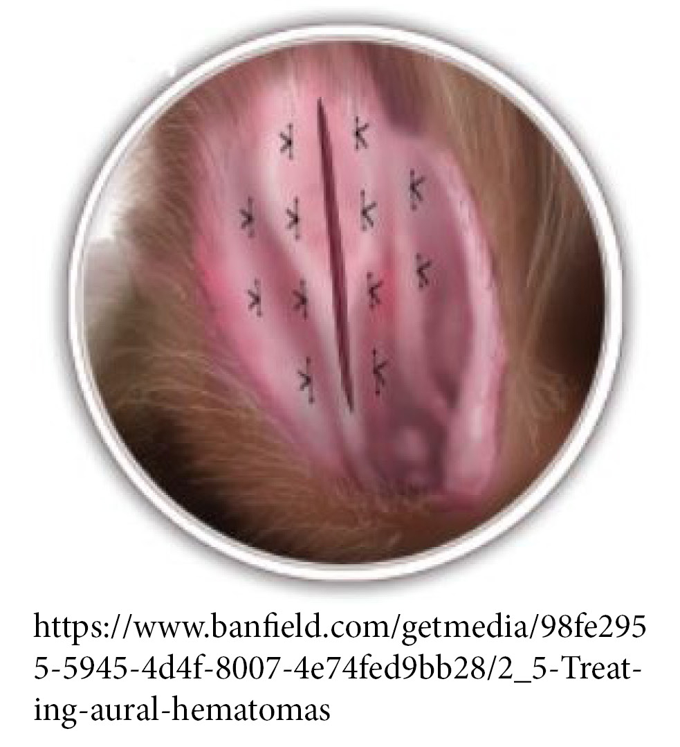 The most successful form of treatment for aural hematomas is surgery to drain out the blood. During this procedure, the incision needs to be left open so that if the ear starts bleeding again, the blood can continue to drain out (otherwise we would have to go in several more times to drain the re-occurring hematoma). To do that, we place several sutures to close the space created by the hematoma, but leave the initial incision open. When the surgery is done, it should look like the illustration on the right.
Since we have seen so many cases recently, I got to watch this surgery for the first time two weeks ago, and then got to assist with another one on Wednesday. This is the first surgery I have performed that is not a spay or neuter, which is exciting!
A crucial part of treating this condition is addressing the primary cause, otherwise aural hematomas will reoccur. Ear infections can be treated with a variety of oral and topical antibiotics, and as I mentioned a few weeks ago, if allergies are at fault, then a change of diet or anti-itch medications (or both) may be in order.
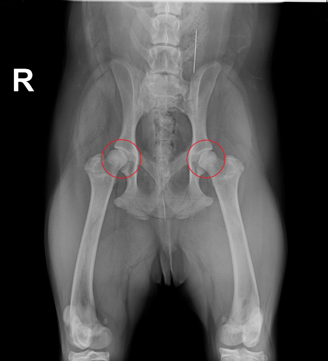 On Thursday, a young dog came in that the owners said had sore hips, was lethargic, and had had some vomiting and diarrhea. We started out by assessing the hips, since he was of a breed predisposed to hip dysplasia. I got to perform full orthopedic, neurologic, and gait examinations, and agreed – his hips seemed pretty sore. His hips didn’t extend or flex very far on either side, his hind end really “wiggled” when he walked (so that he didn’t have to extend his hips as far), and he dragged his toes on the ground with each step. The owners agreed that it was best to take radiographs (x-rays) so that we could get a better picture of what was going on.
Let’s just zoom in a little closer to get a better look…
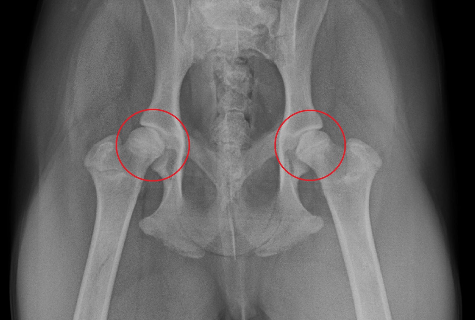
Yep, looks like hip dysplasia to me! The head of the femur (ball) and acetabulum (socket) are not round, and don’t match up nicely with each other the way you’d expect. I don’t see much arthritis yet, in the form of bone remodelling in and around the joint, but this dog isn’t finished growing, so we wouldn’t necessarily expect to see that yet. I start thinking up all of the management and treatment plans we’ve talked about in class in preparation for our discussion with the owner about the diagnosis… but wait a second, didn’t I see…
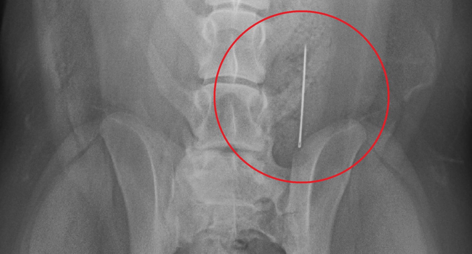
Yuuuuuup, that’s a sewing needle!
Clearly this x-ray was the “money shot” – we solved the sore hips, lethargy, vomiting, and diarrhea all in one go! Thankfully this story has a happy ending. Most foreign objects that make it this far (the large intestine) exit without any troubles. We took a second radiograph using a lateral (side) view to confirm that this sewing needle was in fact encased in a fecal ball, and sent this boy home with strict instructions for his owners to go digging for treasure. The sewing needle passed out uneventfully by the end of the day.
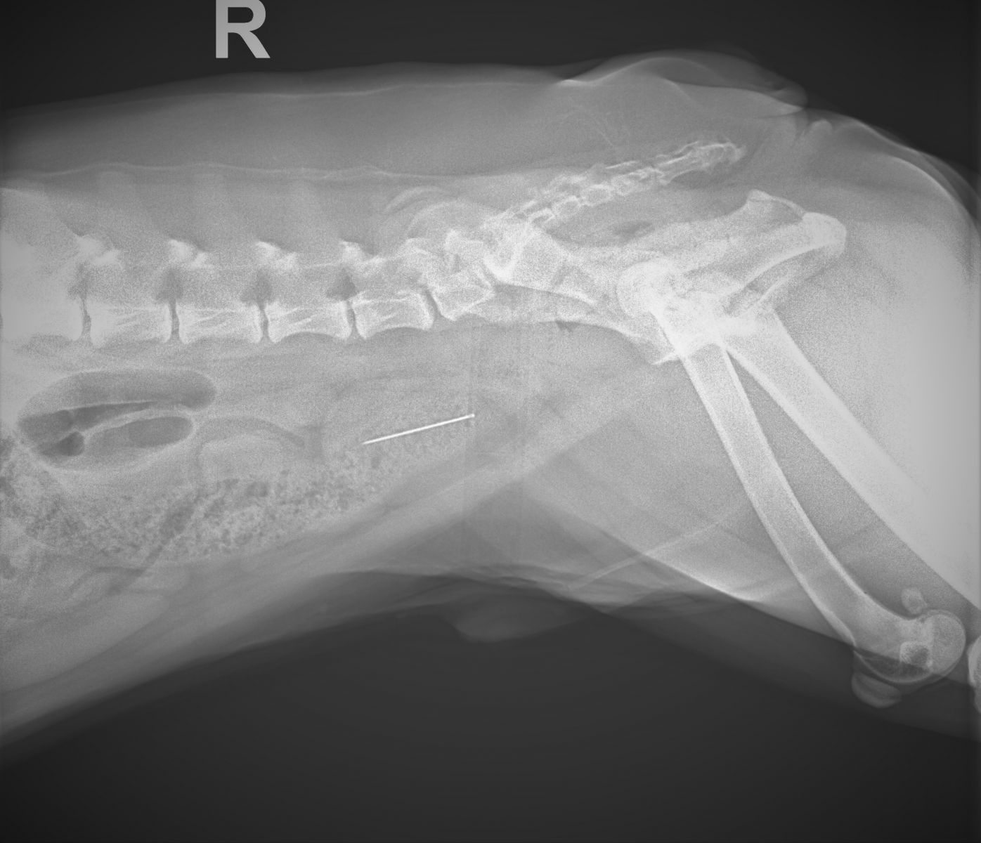
The lateral view showing the sewing needle encased in feces within the large intestine
That’s all I have for week five, I hope everyone has a great week, and make sure to keep your sewing kits nice and high up in the linen closet!