In my first weeks of my externship I was presented with Lily, a sweet 13-year young golden retriever owned by one of the new technicians at our clinic. Lily had a history of scratching her left ear repetitively which resulted in a big bulge on her ear known as an aural hematoma (aural = ear; hematoma = swelling of clotted blood within tissue). Luckily for me, this is a relatively simple surgery that is perfect for a vet student to tackle. Plus, who can resist the opportunity to cut into a fluid filled pouch and squeeze out whatever goop hides inside?
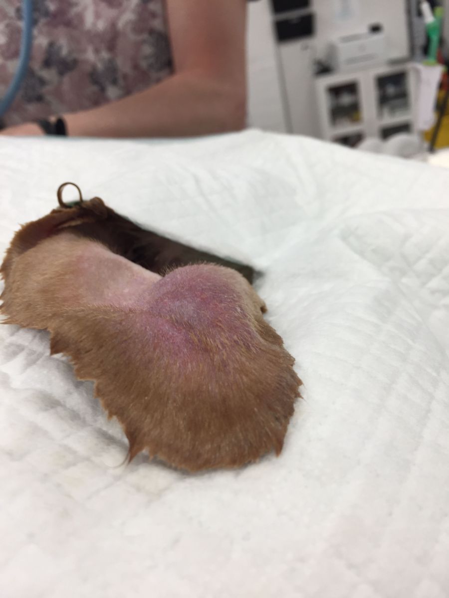 Aural hematomas develop from a dog constantly hitting their ear which then causes an internal bleed between the layers of cartilage in the external ear. This usually is the result of an itchy ear infection or ear irritation which causes the dog to repetitively itch its ear with its paw. The bleeding results in increased pressure which eventually pushes the layers of cartilage apart, forming a blood filled pocket in-between. The pressurized ear may cause even more irritation causing the dog to continue scratching at the ear and making the hematoma worse. Hematomas in the body generally can heal on their own as most surrounding tissues can resorb the clotted blood and shrink the bulge. Due to the nature of the ear’s tissue, an aural hematoma will not heal on its own since the ear cartilage cannot resorb the clotted blood, making surgery the necessary treatment.
The goal of the surgery is to completely open and drain the hematoma and then tack the layers of cartilage together so that they can adhere and heal together. Since this surgery isn’t very invasive, Lily was sedated then maintained on a gas mask. After the ear was clipped and prepared for the surgery I nervously yet excitedly cut a serpentine (curved) incision into the inner surface of the ear, which 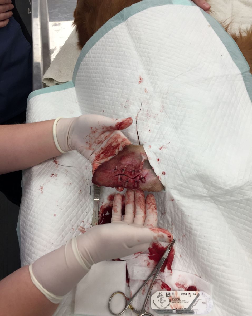 resulted in a satisfying oozing of the blood clot as it was released from its pressurized sac. After pretending to be Dr. Pimple Popper and expressing all of the retained blood clot, I tacked the layers of cartilage together with suture and stents while keeping the incision open to allow for drainage. The next most important part was to bandage the ear to keep it clean and infection free, as well as safe from scratching paws, which is why you’ll see Lily with a pretty hefty bandage. Luckily for her, I had time to decorate it!
Lily came in a couple weeks later after removing the bandage and unfortunately the hematoma was back! Some of my sutures failed allowing for the cartilage layers to stay separated while the incision healed over resulting in a relapsing hematoma. In addition, Lily had managed to scratch through her bandages which continued to irritate the ear. Kindly enough, the owner gave me a second chance to perform the surgery and I was able to improve my techniques to ensure for a successful outcome. Weeks 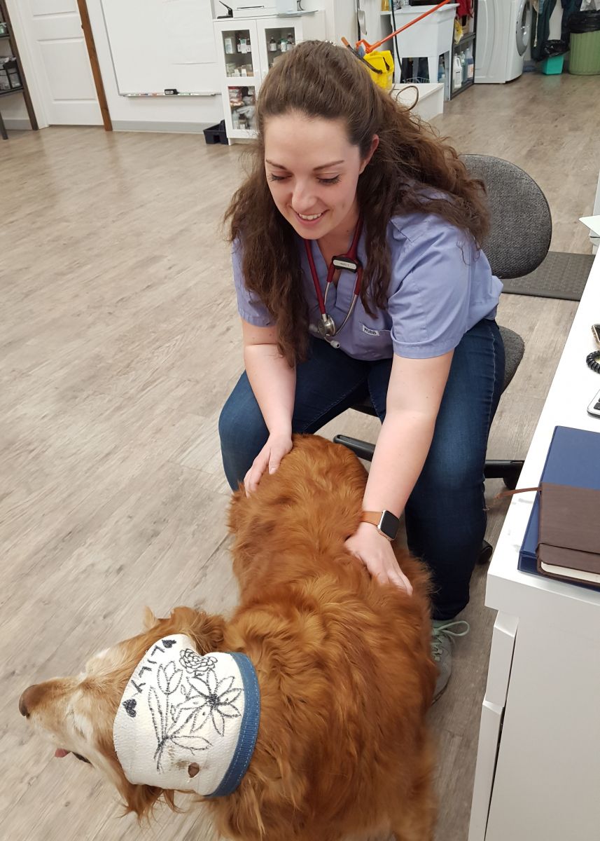 later, Lily’s ear was almost completely healed, and she was relieved to finally have the bandages and cone-of-shame removed for good.
This was the beginning of my surgical adventures with my externship, and I can say that I truly loved holding the scalpel for many more exciting surgeries. From the cow C-sections and dog “lump-ectomies” to removing many testicles and ovaries, I think I might have caught the surgery bug!